Thoracic Outlet Syndrome (TOS)
Along with reading our therapy treatment solution in section 1 below, we also encourage you to read the other 6 sections to fully understand the complexity of this debilitating and potentially dangerous nerve compression syndrome.
(Tap or click to scroll to section)
Section 1: Thoracic Outlet Syndrome (TOS) Therapy Treatment Solution
Section 2: Understanding The Complexities of Thoracic Outlet Syndrome
Section 3: Understanding Muscular Fascia
Section 4: Myofascial Adhesions: Source of the Problem
Section 5: Causes of Thoracic Outlet Syndrome
Section 6: Our Treatment Technologies
Section 7: Your Next Step
Section 1: Thoracic Outlet Syndrome (TOS) Therapy Treatment Solution
Since Thoracic outlet syndrome is the crushing of the neurovascular bundle by the formation of myofascial adhesions, our goals in treatment is to disintegrate all of them thus freeing up the nerves and blood vessels.
We have learned over the past 20 years that the only effective and permanent therapy treatment solution to breaking apart problematic adhesions is with the use of shockwave therapy, the same technology used to break apart kidney stones.
Its powerful acoustic waves quickly disintegrate the adhesion’s collagen bonds involved in the cross-bridged adhered fascial fibres strangling the neurovascular bundle.
However, we don’t’ just use any ordinary version of the technology, but the worlds’ best from EMS (Germany), originators of the technology more than 40 years ago.
Treatment Begins
A gel is applied to the skin over the adhesions , located with precision by our founders incredible sensitive touch being blind. Agun-like applicator depressed against the skin then begins producing the acoustic waves required to reach deep into the hardened adhesions and begin to break them apart.
Sessions start with low-intensity acoustic waves to acclimate patients to the process, start breaking apart superficial fibers in the adhesions, then gradually increase in intensity and depth as treatment progresses.
We must treat every adhesion in the muscles in the neck, chest, along and deep to the clavicle, under-arm, and upper arm, to release the entire neurovascular bundle.
We systematically keep moving the applicator from adhesion to adhesion ensuring all are eliminated.
We continuously monitor your responses and adjust the treatment parameters accordingly to ensure optimal outcomes.
Throughout the sessions, you will feel slight pressure in the muscles as adhesions are being broken apart, accompanied by immediate relief of symptoms caused by that particular adhesion.
You will experience re-creation of symptoms when we begin to break apart an adhesion responsible for those symptoms. For example, you will experience pins/needles sensations in your hand as the adhesion responsible for these symptoms is being disintegrated. Then within minutes you feel the enormous relief to your hand as the nerve no longer causes those sensations. It is an amazing experience.
We prioritize treatments to the side of the body afflicted the worst, to quickly remove dangerous compression from the neurovascular bundle, preventing permanent damage from occurring to its delicate nerve fibers and blood vessels.
Typically we require 3 treatments to remove problematic adhesions along the neurovascular bundle in most cases, but may require 5-7 treatments in severe cases.
We may also decide to begin treating root causes along with the TOS if they are deemed to interfere with our treatments process. For example, we may begin a treatment protocol for neck arthritis while addressing the adhesions it has caused in TOS.
Post-Treatment Effects
You will experience:
Swelling in the arm or hand if present is eliminated by opening up crushed lymphatic channels to allow the accumulation of lymphatic fluid to drain upward to the lymph nodes in the underarm and neck region.
Swelling caused by restricted flow in veins is eliminated whenever the treatments release the crushing of the veins higher up in the neck and chest.
Cold, clammy, pale, and numbness in the hands if present is eliminated by opening up restricted arteries higher up in the neck and chest regions.
Achy elbows, forearms, knuckles on the hand, or anywhere else in the arm is eliminated as the crush forces on the nerves is eliminated.
You can expect skin tenderness, bruising, and swelling for up to 4 days following the first treatment. This is an essential part of the treatment process as we want the body to respond by swelling so the broken collagen proteins can be removed from the treated region and the tissue regeneration phase may then begin.
Every subsequent treatment thereafter will see far less bruising, less swelling, and no skin tenderness. This is because only adhered tissue sees these reactions, healthy tissue sees none.
You can continue with your normal daily activities as the treatments do not require any modification to your normal schedule.
The most amazing aspect to treatments is with your symptoms. You will see an astonishing and in some cases an unbelievable decrease in symptoms immediately. Our EMS shockwave therapy technology is not a toy. It is the original, most powerful in the world so has no problem getting the job done quickly.
Time and Duration
Initial treatments target only 1 side of the neck, chest, clavicular fascia and the arm down to the fingers requiring up to 75 minutes to complete.
If thoracic outlet syndrome is determined to be on both sides of the body, we target the worst side first.
The second treatment will either continue on the original side, depending on how severe the adhesion formation was, or begin the other side in a full treatment there.
A total of 3-5 treatments is typically required to resolve most presentations of thoracic outlet syndrome. Treatments are usually weekly, but some want to slow down the process for logistic reasons and will be treated every 2 or 3 weeks. There is no rush to treat this condition unless your particular case is endangering the neurovascular bundle in which case we would recommend weekly or bi-weekly treatments.
Root Causes
Thoracic outlet syndrome is a secondary condition that developed due to a major, primary root cause, that exposed the neck/chest/shoulder/upper back to excessive and repetitive forces over many years.
Many who suffer from thoracic outlet syndrome are found to have arthritis in the neck, shoulder tendinopathies, pelvic/leg issues…
Once we have addressed the neurovascular bundle compression, we then change course of action and fix the root causes halting any future recurrence of the condition.
However, as mentioned earlier, we may decide to begin addressing causes while addressing the adhesion removal because of how much influence these causes have on the adhesion formation process. We do not want to remove adhesions only to see a particular cause begin to reform them.
Note of Interest
We often encounter individuals who have undergone surgery to remove the first rib in an attempt to alleviate Thoracic Outlet Syndrome. While our therapy treatment solution could have resolved these cases, we still accept these individuals for treatment. It’s important to note that surgery does not address the root causes or underlying adhesions within the scalene muscles and other affected tissues, necessitating our therapy to achieve comprehensive resolution. Our approach offers a non-invasive alternative that targets the root cause of the condition, providing relief and empowerment to those suffering from Thoracic Outlet Syndrome.
Through our innovative therapy treatment solution, we have empowered countless individuals to break free from the grip of adhesions, enabling a life without pain and suffering. The profound transformations experienced by our patients underscore the effectiveness and impact of our approach, paving the way for a future filled with relief and vitality.
Section 2: Understanding The Complexities of Thoracic Outlet Syndrome
Thoracic Outlet Syndrome (TOS) is a condition where the nerves, arteries, and veins—collectively known as the neurovascular bundle—are compressed or stretched by damaged muscles and/or ligaments along their pathway from the neck to the upper arm. This interference can occur anywhere along the path taken by the neurovascular bundle’s origin in the neck to its destination in the fingers. There are several key causes for the involved muscles to suffer damage which will be explained below in section 4.
Anatomy and Pathway of the Neurovascular Bundle
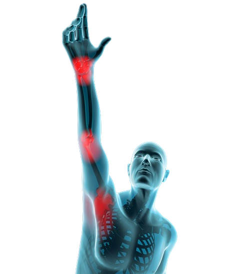
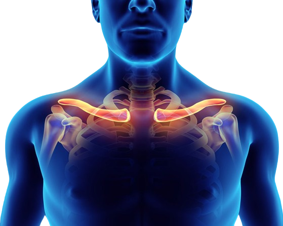
The neurovascular bundle is a complex system comprised of five major peripheral nerves originating from the spinal cord between the fifth cervical vertebra (C5) and the first thoracic vertebra (T1), as well as major arteries, veins, and their numerous smaller branches. These nerves/arteries/veins travel downward along both sides of the neck, pass beneath the clavicles (collarbone), traverse under the chest muscles, and extend into the arms, eventually terminating in the fingers.
The primary muscles involved in Thoracic Outlet Syndrome (TOS) include:
- Scalene Muscles and Related Fascia: Located on the sides of the neck, these three muscles play a crucial role in neck stability and movement
- Pectoral Muscles and Their Fascia: Found in the chest, these muscles aid in shoulder and arm mobility
- Subclavian Muscle and Its Fascia: This is the muscle that lies beneath the clavicle (collar bone) which stabilizes the bone during shoulder motion
These muscles, when healthy with normal elasticity, cover and protect the neurovascular bundle, allowing for the necessary stretching and contracting movements that facilitate neck and shoulder motion. Blood vessels, including arteries and veins, accompany the nerves and receive similar muscular protection, ensuring proper blood flow to the muscles and their nerves.
The key to understanding thoracic outlet syndrome, is to understand muscle health and the fascia every muscle is comprised of.

Section 3: Understanding Muscular Fascia
Fascia is an extremely robust yet pliable type of connective tissue, predominantly comprised of collagen and elastin fibers, which intricately envelop and safeguard every living muscle and nerve fiber (cell) within the body’s muscular framework.
While collagen provides the necessary strength to shield and safeguard these sensitive living muscle and nerve cells, elastin confers the unique elastic capability to stretch and contract, permitting the vital stretching/contracting ability every muscle and nerve requires.
The prefix “Myo” pertains to the fascia associated with muscles, deriving from the Greek term for muscle. Remarkably, a single muscle may harbor upwards of 500,000 living muscle cells, each ensconced within a protective fascial covering known as endomysium.
Furthermore, clusters of these fascia-wrapped muscle cells, numbering around 20,000 in each bundle, are encased by another layer of fascia termed epimysium. Finally, the entire muscle ensemble is enveloped by a third type of fascia called perimysium, which confers upon each muscle its distinct shape. At all three levels, fascia serves the fundamental purpose of binding and safeguarding the living muscle cells.
Crucially, muscular fascia boasts excellent blood flow, facilitated by the living muscle cells’ vascular network, which supplies essential nutrients to the collagen and elastin fibers within the fascia. This intricate system ensures that muscular fascia is adept at absorbing the forces exerted upon it when muscles contract or stretch during joint movement, and the work all muscles allow us to accomplish.
Section 4: Myofascial Adhesions—The Source of the Problem
Myofascial adhesions, commonly known as adhesions, manifest as regions of severely hardened and adhered fascial fibers amidst otherwise healthy elastic fascial tissue.
Their ratio of collagen and elastin changes, resulting in more inflexible collagen fibers and fewer elastic, pliable elastin fibers. The result is a phenomenon where normally straight, healthy muscle fibers see an influx of new collagen fibers that twist and bind other fibers together like glue, forming an intricate weave of cross-bridged collagen fibers, effectively changing the muscle’s tissue structure.
These adhered fascial fibers form an intricate weave of dysfunctional, inelastic, fibrotic, and ischemic bands or large regions within the muscle’s fascial connective tissue layers and even throughout the entire muscle. The term adhesion (adhesive), commonly associated with glue, aptly describes this phenomenon.
Adhesions typically form in the fascial tissue layers due to excessive and repetitive strain activity on its muscular fibers. The adhering and cross-bridged collagen fiber influx is the body’s unique method of protecting the living muscle cellular structures it encompasses from experiencing damage, as living muscle cells can die, resulting in muscle wasting and atrophy.
The result of all this hardening, gluing, twisting, and deformation of a muscle causes:
- The entire muscle to shorten, severely crushing free nerve endings within the muscle, resulting in localized pain
- Decreased healthy oxygen flow into the muscle, causing pain
- Decreased outflow of metabolic waste, causing pain
- The entire muscle to shorten, crushing underlying nerves, arteries, and veins (Thoracic Outlet Syndrome)
Adhesions: Understanding the Consequences of Overstrain
Muscles are marvels of biomechanical engineering, capable of generating significant force to facilitate the myriad activities of daily life. However, this remarkable capacity comes with a caveat: muscles require time to recuperate from the stresses placed upon their fascial network, which serves as vital protectors of the living muscle cells. Given adequate time, fascia can indeed heal from the strains induced by vigorous work activity. The crux of the issue lies in the time factor; frequently, we fail to afford our muscles and their fascia the necessary recovery period amidst the demands of daily living.
Fortunately, the human body possesses an innate resilience, designed to safeguard every living muscle cell under duress. When myofascial tissue is deprived of sufficient recovery time, the body initiates a compensatory mechanism, generating new collagen fibers that intertwine individual fascial fibers in a cross-bridged pattern. Analogous to the reinforcement of a rope, wherein a thicker cord is inherently stronger, this process fortifies the fascial network by binding individual muscle cells and their fascial coverings together, thereby enhancing overall structural integrity.
These cross-bridged collagen fibers effectively act as natural adhesives, binding groups of individual fascial fibers together to form a cohesive, robust unit, thus shielding the strained fibers from further damage. The term “adhesion (adhesive),” commonly associated with glue, aptly describes this phenomenon, wherein fascial fibers become bound together, reinforcing the tissue against the rigors of overexertion.
Consequences of Adhesion Formation & Thoracic Outlet Syndrome
The formation of adhesions initiates a cascade of consequences, as these fibrous connections begin to compress the arteries, veins, and free nerve endings within the layers of fascia surrounding muscles and the neurovascular bundle that lies beneath.
Consequently, a persistent low-grade pain manifests whenever muscles ensnared by adhered fascia are engaged. Moreover, these adhesions have the propensity to shorten certain bands of fascial fibers, exacerbating discomfort upon contraction or stretching of the affected muscles.
With thoracic outlet syndrome, the genesis of repetitive strain adhesion formation can be attributed to a myriad of activities, including:
- Prolonged periods of computer usage with poor ergonomics
- Sustained tilting of the head to view a phone screen
- Engaging in sports without proper muscular conditioning
- Strain imposed by poor posture
- Emotional stress
- Breast enlargement
- Post-breast cancer surgery
- Heavy chest exercising with weights without balancing the back muscles
- Neck arthritis
- Chronic low back pain
- Repetitive cycling over long distances
- Chronic headaches
- Excessive cell phone usage

Section 5: Causes of Thoracic Outlet Syndrome
The scalene, pectoral, and subclavius muscles and their related fascia are the known causes of all forms of thoracic outlet syndrome. What is not commonly known is that these muscles develop damaged, adhered fibers (adhesions) for various reasons.
We have seen and treated thousands of people who suffer from thoracic outlet syndrome and have compiled a list of all root causes we have encountered during our detailed consultations. We have treated patients whose thoracic outlet syndrome has been caused by the following:
- Posture issues: Prolonged forward head posture, rounded shoulders, and poor sitting or standing posture can lead to muscle imbalances, overstrain, and adhesions in the neck, chest, and shoulder muscles, which compress the neurovascular bundle.
- Repetitive strain injuries (RSIs): Frequent, repetitive activities such as typing, writing, lifting, or overhead work can lead to muscular overstrain, adhesions, and subsequent TOS development.
- Trauma: Whiplash injuries, fractures, or other traumatic events involving the neck, shoulder, or upper chest can damage the muscles, leading to adhesions and TOS.
- Sports injuries: Certain sports that involve repetitive arm movements, such as swimming, baseball, or weightlifting, can overstrain the muscles and cause adhesions, leading to TOS.
- Genetic predisposition: Some individuals may have anatomical variations, such as an extra cervical rib, which can increase the likelihood of developing TOS.
- Obesity: Excess body weight can strain the muscles, leading to posture issues and increased risk of developing TOS.
- Pregnancy: Hormonal changes and weight gain during pregnancy can contribute to postural changes and muscle imbalances, potentially leading to TOS.
- Emotional stress: Chronic stress can lead to muscle tension and tightness, increasing the risk of adhesions and TOS.
- Poor ergonomics: Inadequate workstation setup, including incorrect chair height, desk positioning, and monitor placement, can contribute to poor posture, muscle strain, and TOS.
- Age-related changes: Natural aging processes can lead to muscle weakness, reduced flexibility, and increased risk of developing adhesions and TOS.
Section 6: Our Treatment Technologies
Extracorporeal shockwave therapy, initially developed in the 1970s to fragment kidney stones in a process called lithotripsy has evolved over the past 50 years.
We utilize a unique version of extracorporeal shockwave therapy technology (ESWT), first developed by EMS Germany, originators of the technology over 40 years ago, to solve thoracic outlet syndrome.
It still produces the most effective and deep penetrating impulses among all acoustic shockwave generators worldwide by propelling a metal projectile with high pressure compressed air through an exceptionally long cylinder within the applicator to strike an alloy tip. These acoustic (sound wave) impulses are transmitted into the body through a conductive gel on the skin. They create a distinctively high-energy acoustic wavefront, triggering rapid expansion of blood/lymphatic gas molecules, inducing a cavitation vacuum effect within intricately interlinked fascial fibres, succeeded by a swift cavitational implosion force.
This rapid expansion and implosion wave cycle (shockwaves) results in the creation of beneficial fascial adhesion tissue disruption effectively degrading the collagen bonds of adhered fibres, causing them to disintegrate. Physiologically, healthy tissues such as nerves, smooth muscle of organs, outer fascial layers, ligaments, tendons, blood vessels, and lymphatic channels possess varying degrees of elasticity and can expand without repercussions as shockwaves traverse their cellular structures.
Note
EMS researched and thus engineered the first shockwave therapy devices back in the 1980’s for adhesion removal therapy, patented their invention, and to this day still produce the most effective version of the technology. We have no problem breaking apart adhesions or calcium spurring anywhere in the body.
Section 7: Your Next Step
Your journey towards relief begins with taking the next step. Here’s what you can do to initiate the process:
Contact Us:
Reach out to us to schedule a consultation and initial treatment session. Whether you’re local or from afar, we’re here to assist you in finding the best solution for your condition.
We encourage calling us from a land-line phone or e-mail. Cell phones go through dead zones and often our voice message system fails to hear the entire message including a return phone number.
Consultation and Treatment Plan:
During your consultation, we’ll discuss your specific situation in detail and work together to formulate a personalized treatment plan tailored to your needs.
Since treatments require intervals between sessions, we’ll address the logistics to ensure seamless administration of treatments.
Flexible Treatment Options:
Treatments can be administered at any time, even during flare-up situations. Our initial treatment is designed to swiftly reduce painful causes, providing immediate relief.
There’s no “bad” time to begin treatments. The sooner you start, the sooner you’ll experience resolution and regain control over your life.
For more information on our treatment for this condition, or on any other chronic pain condition you may suffer from, we encourage you to contact us to discuss options available to you.

