Iliotibial Band Contracture
Before delving into our innovative therapy treatment solution, it’s essential for you to understand the complexities of this debilitating condition. This comprehensive document aims to address any questions or concerns you may have regarding our highly effective non-surgical therapy solution for Iliotibial Band Contracture.
Definition: What is Iliotibial (IT) Band contracture?
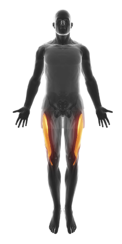
Iliotibial band contracture is a condition where the iliotibial band, a large region of connective tissue on the outer aspect of the thigh, has become dysfunctional. Its fibres have developed lumps and/or bands of twisted tangled fascial fibres that collectively result in an overall shortened (contractured) structure.
We have a 97% success rate in permanently solving iliotibial band contracture because the band’s core fascial tissue structure and resulting contracture process is exactly what we have specialized in treating, not just in the iliotibial band but throughout the body for almost 20 years.
Section Overview:
Section 1: Understanding the Complexities of Iliotibial Band Contracture
Section 2: Understanding Fascia and Fascial Adhesions – Their Role in Iliotibial Band Contracture
Section 3: Causes of Iliotibial Band Contracture
Section 4: Our Treatment Technologies
Section 5: Our Therapy Treatment Solution
Section 6: Your Next Step
Section 1: Understanding the Complexities of Iliotibial Band Contracture
The iliotibial band is a long region of extremely dense and fibrous connective tissue called fascia, directly attached to the outside of the femur. Its role is to stabilize the bone and prevent it from bowing outward during load-bearing activities. This provides the long femur with greater strength, reducing the likelihood of it bending over time, which could lead to a permanent bow shape and eventual bone weakness.
In a male who is 5 feet 8 inches (173 cm) tall, the band is approximately 3 inches (8 cm) deep from the skin of the thigh inward to the bone and nearly 4 inches (10 cm) wide along the entire length of the femur, from hip to knee. The iliotibial band is composed primarily of strong and durable collagen connective tissue fibers, which protect and give every muscle in the body its unique tensile strength and shape.
These extremely strong and inelastic fascial fibers are interwoven into a striated, mesh-like structure, giving the band its shape and strength.

A second type of connective tissue fiber, called elastin, is also embedded within the iliotibial band’s internal matrix, providing slight elastic resistance, similar to a thick elastic band. Without elastin, the inflexible collagen fibers would tear, even with normal daily activities.The iliotibial band is constantly exposed to bowing forces from the femur as we walk, lift objects, climb, jump, and engage in daily activities. The collagen fibers comprising the band are designed to resist normal forces and repair themselves during sleep, preparing for the next day’s activities.
However, there are three distinct problems that initiate the contracture process in iliotibial band contracture:
- Insufficient healing time after activities have caused repetitive excessive strain on the band’s fascial fibers.
- Exposure of the femur to periods of extreme and potentially damaging forces it is not designed to absorb (see Section 3: Causes of Iliotibial Band Contracture).
- Trauma as with falls, sports injuries, car accidents, femur fractures…
As a result, the unique collagen bonds of the band, along with hundreds of thousands of myofascial connective tissue fibers, begin to develop micro-tears due to extreme and repetitive forces over days, months, or even years. Low-grade inflammation soon develops, and the body starts to produce extra collagen fibers to reinforce those that have suffered micro-tears, helping them recover.
If the band continues to be exposed to these extreme and repetitive forces, more fibers will fail, causing further micro-tears, more inflammation, and the onset of pain. The incoming collagen fibers, meant to help repair the damaged ones, harden the existing fibers to prevent further tearing of the band.
This influx of new collagen fibers causes the band’s matrix of non-elastic collagen fibers to become unbalanced, resulting in increased rigidity. More micro-tearing, more inflammation, more hardening, and more pain follow. Fascia is highly innervated with sensitive nerve endings, which, when irritated, can cause extreme pain and weakness as a guarding mechanism to prevent the fibers from tearing completely.
The nerve endings embedded within the band now begin to generate and propagate dysfunctional neurological impulses to the spinal cord, resulting in widespread pain and weakness throughout the thigh muscles. Pain, cramping, achiness, and leg weakness become apparent, making it difficult to walk, climb stairs, and perform other activities.
The iliotibial band’s fascial cellular structure becomes swollen, extremely painful, and sitting for long periods becomes excruciating due to cramping and a relentless ache down the side of the leg to the foot.
You now have developed iliotibial band contracture!
Section 2: Understanding Fascia and Fascial Adhesions –
Its Role in Iliotibial Band Contracture
Fascia Basics
Fascia is a robust yet pliable type of connective tissue, primarily composed of strong, inelastic collagen fibers and elastin, a more flexible form of connective tissue. While collagen provides the necessary strength to shield and safeguard the femur, elastin confers the elastic capability needed for the iliotibial band to stretch and contract slightly whenever the femur bows outward.
Fascia benefits from excellent blood flow, and it is rich in nerves and nerve endings, thanks to a vascular and neurological network that supplies essential nutrients to the collagen and elastin fibers within. This intricate system ensures fascia can absorb the forces exerted upon it during physical activity.
Adhesions typically form in fascial tissue due to excessive and repetitive strain on its collagen fibers. The cross-bridged collagen influx is the body’s unique method of protecting these structures. However, the process results in hardened, twisted, and deformed fascia, leading to several problems:
- The entire iliotibial band shortens, severely crushing the free nerve endings within the band, causing pain
- It decreases healthy oxygen flow to the band, slowing the healing process and causing pain
- It interferes with the normal outflow of metabolic waste, causing pain
- The entire band hardens, crushing underlying nerves, arteries, and veins, directly interfering with the fascia’s regeneration

ITC Trauma – Iliotibial Band Contracture – We resolved this!
Adhesions: Understanding the Consequences of Overstrain
Fascia, a marvel of biomechanical engineering, generates significant force to facilitate everyday activities. However, it requires time to recuperate from the stresses placed upon it. Given sufficient recovery time, fascia can heal from the strains induced by vigorous activity. The issue lies in the lack of recovery time; we often fail to give our fascia the time it needs to recover amidst the demands of daily living.
The human body possesses an innate resilience, designed to protect fascia under strain. When myofascial tissue is deprived of enough recovery time, the body compensates by generating new collagen fibers that intertwine individual fascial fibers in a cross-bridged pattern. Similar to how a thicker rope is inherently stronger, this process fortifies the fascial network by binding individual fibers together, enhancing overall structural integrity.
These cross-bridged collagen fibers act as natural adhesives, binding groups of fascial fibers together to form a cohesive, robust unit. This reinforces the tissue against the rigors of overexertion. The term “adhesion,” commonly associated with glue, aptly describes this phenomenon, where fascial fibers become bound together to protect the tissue from further damage.
Consequences of Adhesion Formation in Iliotibial Band Contracture
The formation of adhesions sets off a cascade of consequences. These fibrous connections begin to compress arteries, veins, and free nerve endings within the layers of fascia that comprise the deep and wide iliotibial band. Adhesions can shorten certain fascial bands, exacerbating discomfort when the band is contracted or stretched.
Section 3: Causes of Iliotibial Band Contracture
As briefly mentioned in Section 1, the causes of iliotibial band contracture are numerous and typically develop over time. However, trauma can also lead to the condition, such as a knee strike to the band during hockey, falling sideways onto an object, or fracturing the femur.
The iliotibial band is designed to absorb normal daily forces on its fibers, but many circumstances can initiate the injury process. Consider the forces on the femur in the following situations:
- Being overweight, which causes extreme bowing forces on the femur
- Engaging in impact sports like jogging, hockey, or tennis with insufficient rest to recover and strengthen
- Working in construction, carrying heavy materials
- Weightlifting with heavy squats (hundreds of pounds) without adequate time for recovery and strengthening
- Being extremely tall with long, thin femur bones, and a normal-sized, thin iliotibial band to stabilize it
- Experiencing excessive forces from an antalgic gait due to a degenerating knee or hip joint
- Sustaining trauma, such as femoral bone fractures requiring plates, pins, or screws
- Falling on the side of the thigh
- Developing lumbar spine arthritis, which causes prolonged excessive forces on the band
- Adductor (groin) injuries
Another major factor to consider is pelvic alignment. If the pelvis is not in a neutral position, the large tensor fascia latae (TFL) hip flexor muscle can develop myofascial adhesions (see Section 2: Myofascial Adhesions). This causes the TFL to shorten and place direct stress on the iliotibial band since they are part of the same structure. The TFL provides tension for the band, so if it becomes excessively tight due to pelvic posture, the iliotibial band will also tighten.
If the adductor (groin) muscles are tight, the femur will internally rotate, increasing tension in the TFL due to the formation of myofascial adhesions, which in turn causes contracturing forces on the iliotibial band. As you can see, the iliotibial band can bear excessive forces due to abnormal pelvic posture, hip flexor contracture, or direct trauma to the thigh, all of which can negatively affect the surrounding muscles and connective tissues.
Section 4: Our Treatment Technologies
Extracorporeal shockwave therapy (ESWT), initially developed in the 1970s to fragment kidney stones in a process called lithotripsy, has evolved significantly over the past 50 years.
We utilize a unique version of ESWT, first developed by EMS Germany, the originators of the technology, to treat iliotibial band contracture. It produces the most effective and deep-penetrating impulses among all acoustic shockwave generators worldwide. This is achieved by propelling a metal projectile with high-pressure compressed air through an exceptionally long cylinder within the applicator, which strikes an alloy tip. These acoustic (sound wave) impulses are transmitted into the body via a conductive gel applied to the skin.
The impulses create a distinctive high-energy acoustic wavefront, triggering the rapid expansion of blood and lymphatic gas molecules, inducing a cavitation vacuum effect within the intricately interlinked fascial fibers, followed by a swift cavitational implosion force.
This rapid expansion and implosion wave cycle (shockwaves) results in beneficial fascial adhesion tissue disruption, effectively degrading the collagen bonds of adhered fibers, causing them to disintegrate. Physiologically, healthy tissues such as nerves, smooth muscle of organs, outer fascial layers, ligaments, tendons, blood vessels, and lymphatic channels possess varying degrees of elasticity and can expand without negative repercussions as the shockwaves traverse their cellular structures.
Note:
EMS researched and engineered the first shockwave therapy devices back in the 1980s for adhesion removal therapy, patented their invention, and to this day still produce the most effective version of the technology. We have no difficulty breaking apart adhesions or calcium spurring anywhere in the body.
Section 5: Our Therapy Treatment Solution
Our therapy treatment solution for iliotibial band contracture was one of the first chronic pain conditions we successfully solved 20 years ago. Here’s a comprehensive overview of our therapy treatment protocol.
Now that our diagnostic process has identified all root causes, secondary soft tissue complications, and myofascial adhesion formation regions throughout the body responsible for the development of iliotibial band contracture, we are ready to resolve it.
Since the iliotibial band is comprised entirely of fascia, and the fascia has developed hardened, cross-bridged, damaged regions of collagen fibers, our solution is to break the collagen bonds throughout the entire band using the world’s best shockwave therapy technology. We don’t use just any ordinary version of the technology, but the most effective and deep-penetrating in the world, developed by the originators in Germany, EMS.
Therapy Treatment Parameters
We prioritize treating the sections of the band most affected and causing the most severe symptoms, aiming for a rapid elimination of these symptoms.
Administering Shockwave Therapy
As mentioned in our technologies section, we use EMS shockwave technology to quickly disintegrate the adhesion’s collagen bonds involved in the cross-bridged fascial fibers. A gel is applied to the skin over the region, and a gun-like applicator begins producing the acoustic waves required to reach deep into the hardened adhesions. We have no problem breaking apart adhesions or embedded deposits, which are common in severe cases of iliotibial band contracture.
Treatment Begins
We start with you lying on your side, and the acoustic waves of our EMS technology penetrate the fascial fibers of the band through the applicator placed over your skin. A gel is applied to ensure the acoustic waves enter the band without attenuation.
Sessions start with low-intensity acoustic waves to acclimate you to the process, beginning to break apart superficial fibers in the adhesions, then gradually increase in intensity and depth as treatment progresses.
We continuously monitor your response and adjust the treatment parameters to ensure optimal outcomes.
During the session, you will feel slight pressure in the band as adhesions are being broken apart, accompanied by immediate relief from symptoms caused by that particular adhesion.
Our founder, who is blind, uses his highly developed palpation skills to locate and guide the shockwave application, track the progress of adhesion dissolution, and reposition the applicator as needed to target specific areas.
Treatments typically last between 50 and 75 minutes. We systematically locate the largest, most afflicted regions first and begin disintegrating their collagen bonds in a slow, gentle manner, as the band is extremely sensitive.
As the treatment progresses, you will feel the fascial pressure in the band begin to decrease, the sensitivity lessen, and warmth invade the treated region. Increased circulation and decreased levels of painful metabolic waste chemicals are noticeable within minutes.
There may be tenderness during the treatments, but it will be within normal tolerance, and we adjust the acoustic wave pressure according to your comfort level.
In subsequent treatments, we gradually widen the treated areas to eventually cover the entire iliotibial band, tensor fascia lata muscle, and fascia around the patella (knee cap), as all these structures are involved in the contracture.
By the end of the treatment process, we will have disintegrated every collagen bond that had formed within the deep layers of the band. A total of four or five sessions may be required to fully resolve iliotibial band contracture, each adding more warmth, less pain, and improved leg strength.
Expectations
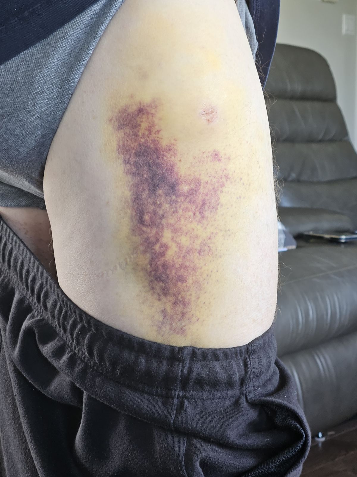
You may experience bruising in the worst regions of the band following the first two treatments. Swelling and tenderness are also part of the process as the body begins to clear out the broken collagen fibers and regenerate the iliotibial band back to its normal health.
Treating the Causes
During the treatments, we also address all the root causes of your iliotibial band contracture. As chronic pain specialists with 20 years of experience, we draw on our vast knowledge to fix issues in the feet, knees, hips, pelvis, and even the neck, if they are found to contribute to your condition. For example, problems with the adductor (groin) muscles can lead to iliotibial band contracture due to the biomechanical imbalance in pelvic alignment.
What to Expect After Treatments
It may take up to two months for your body to fully regenerate the iliotibial band, but you will experience immediate and increasing relief after each treatment.
- All symptoms will gradually improve, including:
- Lessening of thigh pain
- Reduction and eventual elimination of pins-and-needles sensations or numbness in the region
- Increased strength in the entire leg
- Decreased cramping and tension after sitting for extended periods (in cars, chairs, etc.)
- Elimination of low back, knee, and hip pain
- Elimination of abnormal cold or hot sensations
Section 6: Your Next Step
Your journey towards relief from iliotibial band contracture starts with taking the next step. Here’s how to begin:
1. Contact Us:
Reach out to us to schedule a consultation and your initial treatment session. Whether you’re local or coming from afar, we’re here to guide you toward the best solution for your condition.
2. Consultation and Treatment Plan:
During your consultation, we will discuss your specific situation in detail and work together to create a personalized treatment plan tailored to your needs and availability.
Since treatments require intervals between sessions, and most individuals benefit from 3-5 sessions, we will help coordinate logistics to ensure a smooth treatment process.
3. Flexible Treatment Options:
Treatments can be administered at any time, including during flare-ups. Our initial treatment is designed to quickly reduce painful inflammation, offering immediate relief.
There’s no “bad” time to begin treatment. The sooner you start, the sooner you’ll experience relief and regain control over your life.
For more information on our treatment for iliotibial band contracture, thoracic outlet syndrome, or any other chronic pain condition, we encourage you to contact us and explore the options available to you.


